Injectable Depot
Gels at body temperature
Resorbable, biocompatible depot
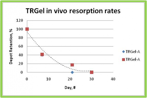
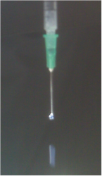
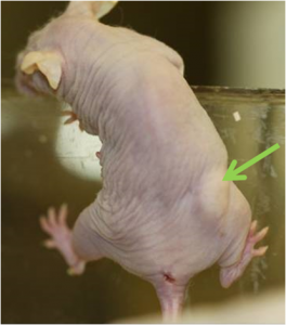
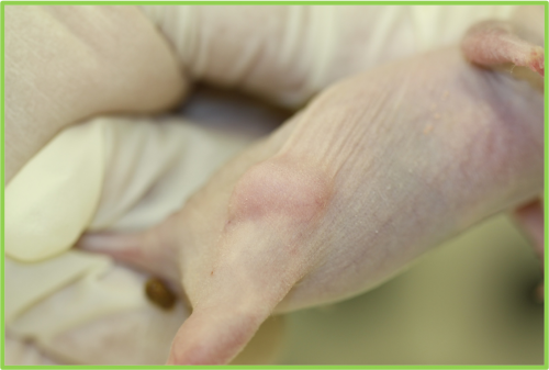
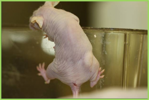
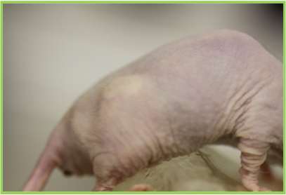
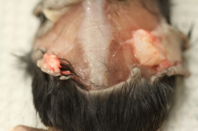
Poly (2-3) glucose T1-30 can be injected subcutaneously by syringe at room temperature. As the polymer solution warms in the body it forms a hydrogel depot which then slowly resorbs. The depot is useful as an envelope to localize emulsions or microcapsules, as a vehicle for drug delivery, as a liquid emobolic, and as a dermal filler. The photo above shows a depot of our T1-30 polymer injected as a 10% wt/v solution in a mouse model. The resorption time is about 60 days.
Envelope Gel
The hydrogel depot is porous for soluble species up to 500 kDa, but it retains larger soluble molecules and insoluble particles. Sufficiently large or insoluble materials that are injected with a poly (2-3) glucose solution will be localized in the hydrogel envelope. For example, a 10 kDa chemokine dissolved in polymer solution and injected, will rapidly diffuse out of the hydrogel, but the same chemokine embedded in 150 um microcapsules will be localized in the depot as the microcapsules elute. Similarly, an emulsion of a lypophilic drug or a suspension of drug crystals will remain localized until the drug gradually dissolves.
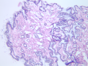
Depots encapsulate many particles. Alternatively, a single cell can be individually encapsulated by a cell encapsulation method . Individual cell encapsulation minimizes the diffusion distance for transport of oxygen and nutrients to the cell. Poly(2-3)glucose is compatible with cells. The murine tissue sample shown above was taken from tissue adjacent to an injected depot. The histology is uniform and healthy.
Liquid Embolic
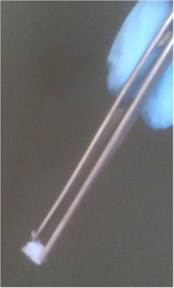
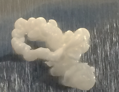
A dense hydrogel can be placed in a blood vessel via catheter to block blood flow in an arteriovenous malformation or aneurysm. While the polymer solution residence time can be low enough to avoid gelling in a short needle, catheter delivery requires gelation suppression. The most direct approach to avoid clogging the catheter is by initially dissolving the polymer in DMSO. Once the solution is extruded into the blood, the DMSO diffuses away and the polymer gels. The video below/left shows poly (2-3) glucose T1-25 polymer in DMSO delivered into PBS by a 1.5 m long catheter. The catheter is at room temperature, and the PBS is at 37 C. The polymer solution rapidly gels when it contacts the PBS. The image below/right shows the recovered, extruded polymer.
Catheter Injection
Poly (2-3) glucose T1-25 dissolved in DMSO and delivered by 533 um x 1.5 m catheter into 37 C PBS.
Liquid Embolic
Dermal Filler
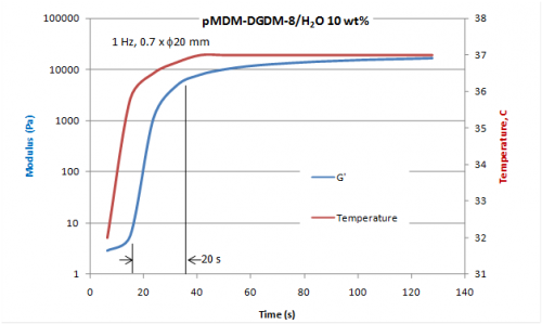
Achieving a natural-looking long-lasting desired aesthetic outcome depends on the rheological properties of the dermal filler. Vicsoelasticity and cohesivity determine resistance of the filler to deformation and they control gel spreading. Typical fillers such as hyaluronic acid gels or collagen must compromise on rheologies that are fluid enough for needle injection yet solid enough to resist deformation and spreading. Thermal reverse gelling poly(2-3) glucose is a low viscosity liquid solution in the needle and an elastic hydrogel at body temperature. Thermal phase change enables much greater resistance to deformation and spreading than single phase fillers. The phase transition from gel to liquid can be used to dissolve an injection error with local cooling of the skin. APIs such as botulinum toxin can be mixed in the poly(2-3) glucose solution and then localized with the injected gel.
Poly(2-3)glucose T1-35 provides long residence time in the needle and good resistance to deformation and spreading. A 10 wt/v% solution can easily pass through a 1 ml syringe with a 27 gauge needle at a plunger speed of 10 cm/min. It is non-toxic and hypoallergenic. Modulus is controlled by polymer solution concentration (2-15 w/v%) prior to injection.
Controlling Resorption Time with Composition
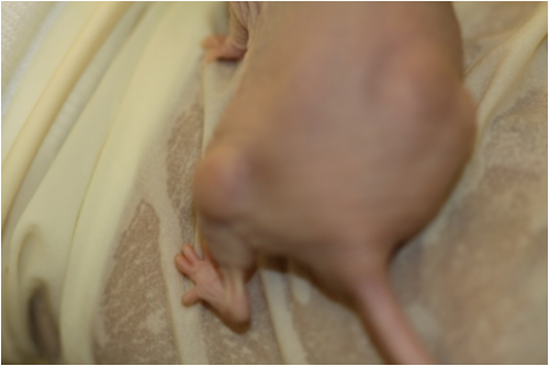
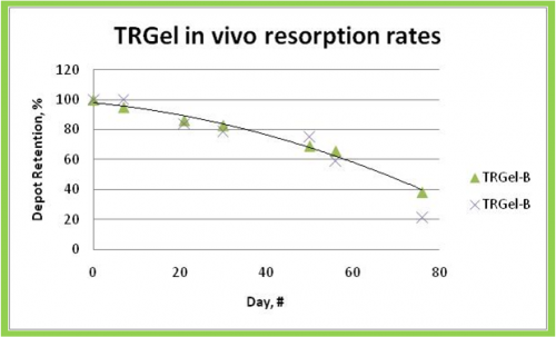
The figures below illustrate resorption rates of two different poly (2-3) glucose compositions. Two mice each received two depots of different composition. Excision at ten days shows the difference in depot size. One composition lost 80% of its volume in 15 days and the other resorbed the same volume in 90 days.
Slow and Fast Resorption
Slow resorption Illustration
Tunable resorption rate
Fast resorption Illustration